- 2026 版正式建議用 externally validated 風險模型辨識高風險 AKI 病人(2C)與預測需要 RRT 的成人(1C)— 但模型必須 externally validated,且要 link 到具體 action(不能孤立 alert)。
- 反對單純 SCr-based interruptive EHR alert(1B)— 多個 RCT 顯示孤立 alert 不改善 outcome 反而增加 alert fatigue。
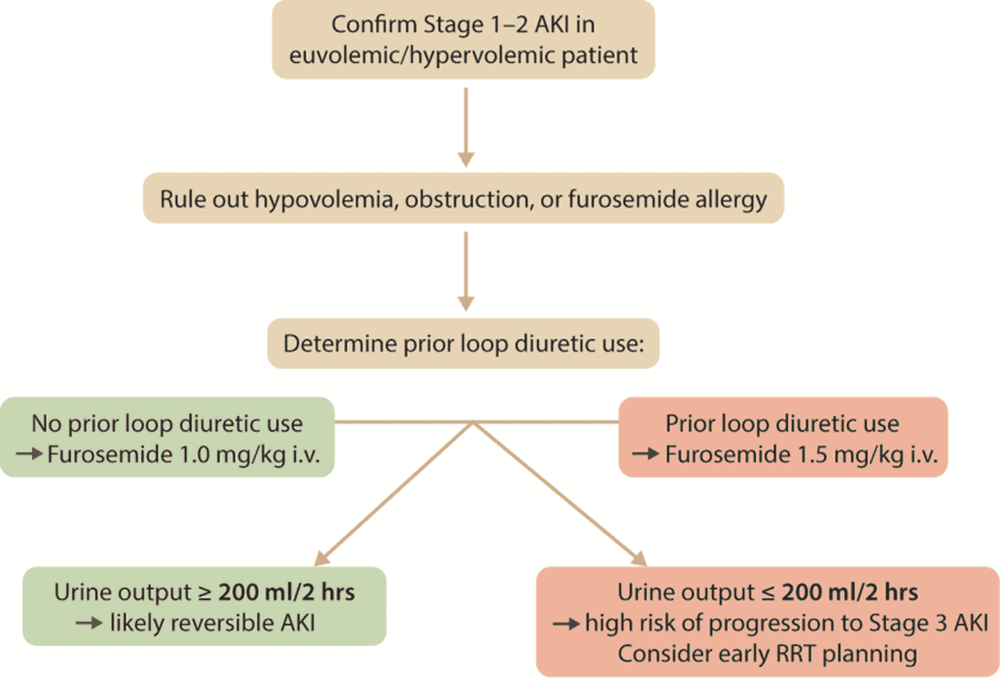
- Furosemide Stress Test (FST) 正式入指引:Stage 1–2 euvolemic/hypervolemic AKI 預測進展風險(成人 2C、兒童 2B)。Figure 7 完整流程。
- Kinetic eGFR (KeGFR) 在 non-steady-state(SCr 變動中)的劑量決策時計算(2B)。
- Damage biomarker(TIMP-2*IGFBP7、NGAL、KIM-1、L-FABP)依其 regulatory approved 適應症使用以早期識別 moderate/severe AKI(成人 2B、兒童 2C)— PrevAKI 與 BigpAK 兩個 RCT 確立其指引價值。
- Urine microscopy(muddy brown casts、RTE cells)在病因不明時做(2D);renal angina index(兒童 ICU)正式入指引(2B)。
2.1 風險預測模型 — 在 AKI 還沒發生前先找出來
核心理念:AKI 一旦發生再處置已經太晚 — 真正的腎保護要在 exposure 之前就鎖定高風險族群。2026 給出兩條建議:
- Recommendation 2.1.1(2C):用 externally validated 臨床風險模型辨識高 AKI 風險的兒童與成人,並指導腎保護策略。
- Recommendation 2.1.2(1C):用 externally validated 風險模型辨識成人 AKI 中可能需要 RRT 者,協助 informed choices 與 shared decision-making。
哪些模型 externally validated?
| 族群 | 模型 | 表現 |
|---|---|---|
| Cardiac surgery 成人 | Mehta score(Cleveland Clinic、AKICS、SRI) | 預測 stage 3/RRT 中度準確(AUROC 0.74–0.82);廣為 external validation |
| Non-cardiac surgery 成人 | SPARK score(Korean cohort 衍生) | 預測 incident AKI 高 certainty 但 accuracy 偏低;stage 3 中度準確 |
| PCI 顯影劑暴露 | Mehran score(含 update 版)、Bartholomew、Tsai 等 | 多項 external validation;Mehran 仍是 cath lab 最常用 |
| Community-acquired AKI(低資源環境) | ISN 0by25 Clinical Risk Score(Table 15) | 10 點簡化工具,適用於缺乏 SCr 設施的地區 |
| Hospital-wide 成人 | 多項 EHR-based AKI prediction models(含 ML 模型) | 本地表現需重新測試;NHS 與多個美國醫療系統已部署 |
| 兒童 ICU | Renal Angina Index (RAI) — 見 §2.6 與 Table 20 | 中度 certainty;預測 severe AKI 與 RRT |
來源:KDIGO 2026 Clinical Practice Guideline for AKI and AKD (Public Review Draft, March 2026) — 個人教學使用,原始版權屬 KDIGO。
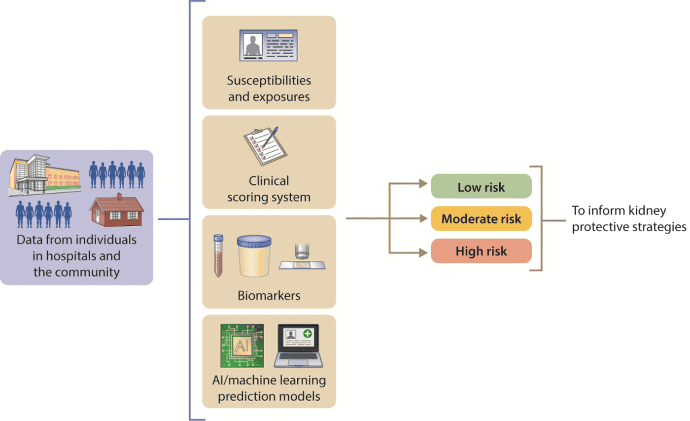
Figure 5 是這章的 conceptual figure — 把 susceptibilities + exposures、clinical scoring、biomarkers、AI/ML models 四個資料來源整合,最後分流出 low / moderate / high risk 三組,對應不同的腎保護策略強度。
來源:KDIGO 2026 Clinical Practice Guideline for AKI and AKD (Public Review Draft, March 2026) — 個人教學使用,原始版權屬 KDIGO。
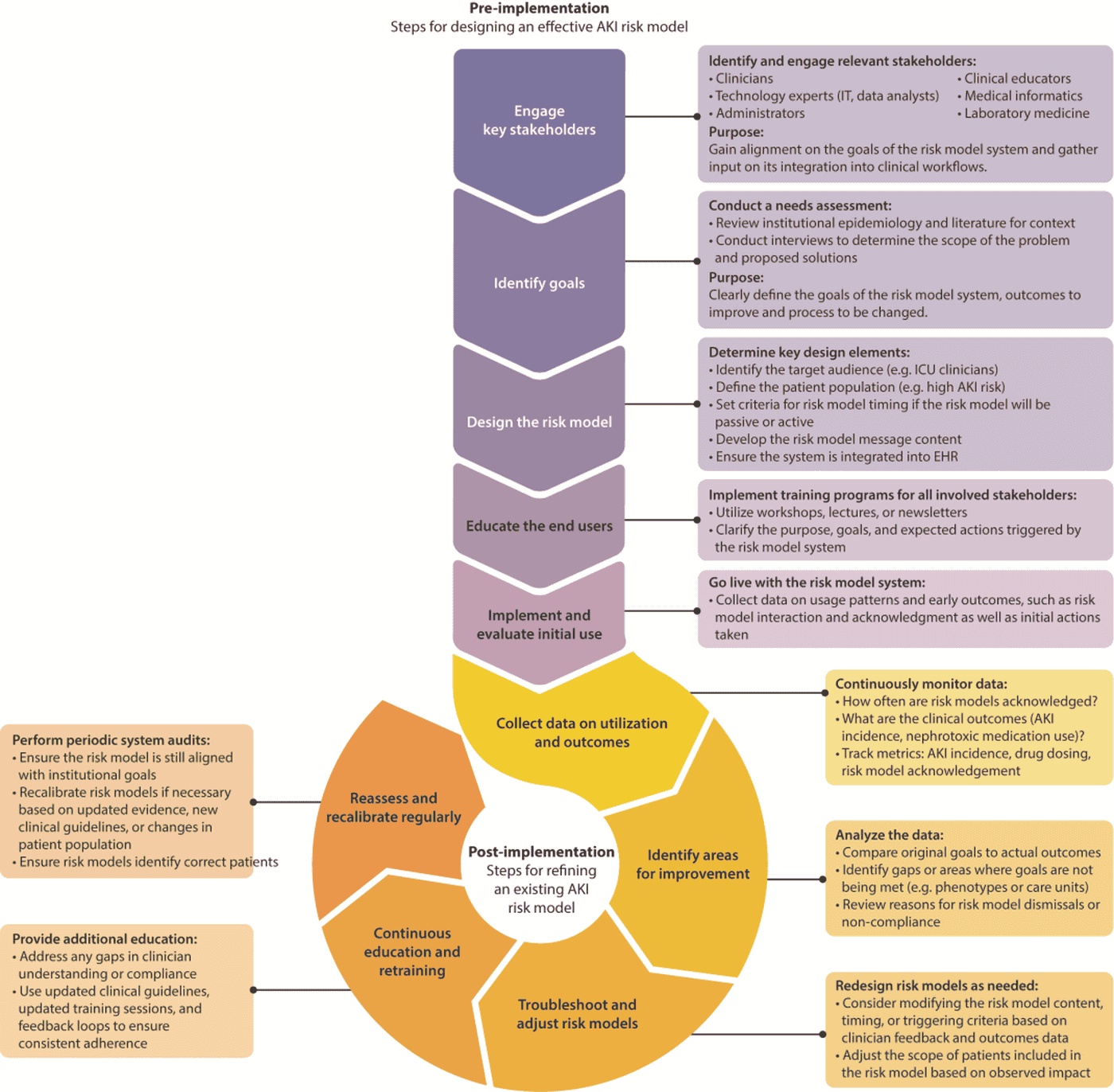
Figure 6 是給 EHR 部署團隊看的 — 預測模型不是「導入就結案」,而是要 pre-implementation(development → validation)+ post-implementation(continuous monitoring → recalibration)的全生命週期管理。這直接呼應 Practice Point 2.1.2:「在哪裡部署就要在哪裡 re-test 性能」。
2.2 EHR 自動警示 — 反對孤立的 SCr alert
Recommendation 2.2.1(1B):反對在沒有 systematic response strategy 的情況下,於成人常規臨床照護中發送 interruptive、creatinine-based 的 AKI EHR alert。
為什麼?多個大型 RCT(Wilson NEJM 2015、Selby JAMA 2019、Tachet du Borgne JAMA 2024)一致顯示 — 孤立的 SCr alert:
- 不改善病人結局(mortality、CKD、RRT 都沒差)
- 但增加 alert fatigue(其他重要 alert 反而被忽略)
- 且 SCr 動力學本身有 delay → alert 跳出來時 AKI 早已發生數小時/天
- 缺 baseline SCr 的時候 alert 易誤判
正確用法:alert 必須與 standardized response pathway 綁定 — 例如 alert 同時觸發 nephrology consult、自動暫停 nephrotoxin、調整劑量、提示 follow-up time stamp 等。「alert + action bundle」這種設計才有實證價值(如 Sutherland UCSF 模式)。
2.3 Kidney functional reserve、Furosemide stress test、KeGFR — 動態評估三劍客
2.3.1 Kidney functional reserve(KFR)
Practice Point 2.3.1:在可能的 exposure 前評估 kidney functional reserve(KFR)可幫助識別高 AKI 風險者。方法見 Table 16:
- Oral protein loading(最生理但變異大)
- Amino acid infusion(standardized 但需 IV)
- Creatinine clearance before / after protein load(需 24h 收集)
Husain-Syed 等人在 elective cardiac surgery 病人發現:所有人靜息 GFR 正常,但 KFR ≤15 ml/min/1.73 m² 的人術後 AKI 風險增 11.8 倍(AUC 0.83,優於 traditional SCr)。HSCT 受贈者同樣顯示 KFR <20% 與後續 persistent kidney dysfunction 風險增 5 倍。
2.3.2 Furosemide Stress Test(FST)
Recommendation 2.3.1:建議在 Stage 1–2 AKI 的 euvolemic 或 hypervolemic 成人(2C)與兒童(2B)使用 FST 預測進展到 Stage 3 AKI(含 RRT)的風險。
來源:KDIGO 2026 Clinical Practice Guideline for AKI and AKD (Public Review Draft, March 2026) — 個人教學使用,原始版權屬 KDIGO。
🧭 圖表逐步解析:Figure 7(FST 應用流程)
這張圖在回答什麼問題:面對一個 Stage 1–2 AKI 病人,會不會進展到 Stage 3 / 需要 RRT?FST 在 2 小時內給你答案,幫你決定是否提早規劃 RRT 或加強腎保護。
逐步走完:
- ≥200 ml/2hr → likely reversible AKI(可逆性高,常規腎保護即可)
- <200 ml/2hr → 進展到 Stage 3 AKI 風險高,提早規劃 RRT、加強監測、跟家屬討論
容易踩雷的地方:
- 對 hypovolemic 病人做 FST。最危險的錯誤 — furosemide 會誘發 hypotension 與後續腎傷害惡化。做之前先用 POCUS 評估 IVC、用 dynamic indices(PPV、SVV、passive leg raising)確認非 fluid-responsive。
- 對長期高劑量 loop diuretic 病人的「No response」誤判為 high-risk。Diuretic resistance 可能掩蓋真實腎儲備力。在這群病人 FST 解讀要保留。
- 2 小時前就決定打第二劑。必須等滿 2 小時觀察 — 提早給多劑會無法解讀。
- 不記錄 baseline 體液狀態。FST 結果 + baseline volume status 才有意義。建議用 standardized FST checklist。
- 把 FST 當「diagnostic test」。它是 prognostic test(預測進展),不是診斷工具。應該與其他 tool 一起使用。
2.3.3 Kinetic eGFR(KeGFR)
Recommendation 2.3.2(2B):在住院成人 AKI 風險或已有 AKI、且 SCr 處於 non-steady state、又需要 GFR 估算來做臨床決策時,計算 kinetic eGFR。
為什麼這重要?傳統 CKD-EPI eGFR 假設 SCr 穩定 — 但 AKI 病人 SCr 每小時都在變,傳統 eGFR 會嚴重高估真實 GFR(病人「真實」GFR 可能 15,但因 SCr 還沒爬上來而算出 60)。KeGFR 把 SCr 變動率納入計算,給出更接近真實的 GFR estimate。臨床意涵:
- 抗生素劑量(vancomycin、aminoglycoside、β-lactam)— 用 KeGFR 可避免過量
- 顯影劑暴露決策 — 真實 GFR <30 vs >30 影響 contrast volume 上限
- 加護升等(CRRT preparation timing)— KeGFR 持續低於 15 ml/min 是 escalation 訊號
2.4 Biomarker — 從研究工具變成臨床決策工具
Recommendation 2.4.1:在 AKI 高風險的 acute care 情境,建議用 kidney tubular stress 或 damage biomarker(依其 clinically validated / regulatory approved 適應症)早期識別 moderate or severe AKI 風險者,以指導 targeted 腎保護策略(成人 2B、兒童 2C)。
關鍵 biomarker 與其臨床用途:
| Biomarker | 來源 | 反映 | 主要 RCT 證據 | 臨床使用 |
|---|---|---|---|---|
| [TIMP-2]*[IGFBP7](NephroCheck) | Urine | Tubular stress(cell cycle arrest) | PrevAKI(2017):cardiac surgery + biomarker >0.3 → KDIGO bundle 介入 → AKI 從 72%→55%。BigpAK(2021):abdominal surgery 類似結果 | ICU 入住 12h 內、major surgery / sepsis 後高風險窗 |
| NGAL(urine 或 plasma) | Urine / blood | Tubular damage(lipocalin family) | 多項 cohort + 部分 RCT;在 cardiac surgery、ICU、HRS 鑑別有用 | 院內高風險、HRS 鑑別 ATI vs HRS 的 adjunct |
| KIM-1 | Urine | Proximal tubule injury | 多項 cohort、無 RCT;mortality 預測中度準確 | 研究階段較多;接受 nephrotoxic 治療者監測 |
| L-FABP | Urine | Tubular ischemia/oxidative stress | 多項 observational;日本廣泛使用 | ICU、cardiac surgery、CKD risk stratification |
| Cystatin C | Serum | Glomerular filtration | 大量 cohort;2026 已入 AKI 診斷準則 | SCr 失準時取代 |
Practice Point 2.4.2:[TIMP-2]*[IGFBP7] 在 ICU 入住 12 小時內或 major surgery / sepsis 後可用於 moderate-severe AKI 風險評估。
Practice Point 2.4.3:urine NGAL 在兒童與成人住院場景識別高 severe AKI 風險者。
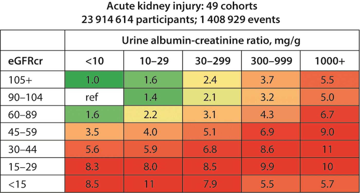
Practice Point 2.4.1(眉角):Baseline albuminuria(UACR 或 dipstick)提供獨立於 eGFR 的 AKI 風險預測 — 心血管門診若已有 UACR 數據,本身就是 AKI 風險工具。
來源:KDIGO 2026 Clinical Practice Guideline for AKI and AKD (Public Review Draft, March 2026) — 個人教學使用,原始版權屬 KDIGO。
Figure 8 是這個 albuminuria-as-risk 概念的視覺化:49 個 cohort、近 2,400 萬人、140 萬 AKI 事件的 pooled analysis。橫軸是 UACR 5 級,縱軸是 eGFR 7 級,網格內數字是 AKI hazard ratio(vs reference 即 eGFR 90–104 + UACR <10)。重點觀察:
- UACR ↑ 在所有 eGFR 級別都會增加 AKI 風險(每個 row 都是右側 redder)
- UACR 1000+ 即使 eGFR ≥90,AKI HR 仍達 5.5 — 跟 eGFR 45–59 + UACR <10(HR 3.5)相當
- 對心臟科:HF + albuminuria(即使 SCr 正常)= AKI 高風險,cath/contrast 前要特別小心
2.5 Etiology 與 Diagnostic Evaluation
2.5.1 Urine microscopy(尿沉渣鏡檢)
Recommendation 2.5.1(2D):成人 AKI 在病因不清楚或懷疑 intrinsic kidney disease 時,建議 urine microscopy 作為 diagnostic evaluation 的一環。
關鍵發現(Practice Points 2.5.1–6):
- Muddy brown granular casts + RTE cells:高度支持 acute tubular injury (ATI),可區分 hemodynamic(prerenal)型;也預測進展到 severe AKI
- Urine eosinophils:低 sensitivity、中度 specificity — 不能用來排除 AIN(過去常用,但實證不支持)
- Urinary inflammatory cytokines:可幫助區分 AIN vs other AKI
- FeNa / FeUrea:不應常規用 — 整體臨床效用有限;只在選定情境(如 cirrhosis-associated AKI 鑑別 HRS)作為 adjunct
- Cirrhosis-associated AKI 鑑別:FeNa <0.1% + plasma/urine NGAL 可協助區分 HRS(low FeNa, low NGAL)vs ATI(low FeNa 但 NGAL 高)
| Urine sediment finding | 對應 syndrome |
|---|---|
| Muddy brown granular casts、RTE cells | Acute tubular injury (ATI) |
| RBC casts + dysmorphic RBC | Glomerulonephritis |
| WBC casts、eosinophils(有限) | AIN(acute interstitial nephritis) |
| Crystalluria | Drug-induced crystalline nephropathy(acyclovir、sulfa、indinavir、methotrexate) |
| Hyaline casts only | Prerenal(hemodynamic)— 但專一性低 |
| Pigmented casts + heme dipstick(+) without RBC | Rhabdomyolysis / hemolysis |
2.5.2 Kidney biopsy 與影像
- Practice Point 2.5.7:unexplained AKI 在臨床評估無法明確分辨病因、或結果會影響管理時,應考慮 kidney biopsy
- Practice Point 2.5.8:Kidney Doppler 超音波(含 renal resistive index, RRI)在重症族群可協助 AKI 進展風險分層 — RRI >0.7 與 persistent AKI 相關
- Practice Point 2.5.9:住院/重症 AKI,POCUS 可幫助評估 volume status 並區分 volume responsive vs intrinsic AKI(IVC、心室填充、肺超音波 B lines)
2.6 兒童專用:Renal Angina Index
Recommendation 2.6.1(2B):對 AKI 高風險的重症兒童,建議用 renal angina index (RAI) 識別 severe AKI 與 RRT 風險者。
RAI 的概念跟 cardiology 的「angina = pre-MI warning」類似 — 在腎傷害發生前用風險 × 早期變化合計成總分(Table 20):
| Risk score(×) | Injury score |
|---|---|
| 1 = ICU 入住 (low risk) 3 = solid organ / BMT 5 = mechanical ventilation + vasoactive | 1 = SCr 上升 < 0.5× baseline(相對 baseline);UO ≥ 1 ml/kg/h 2 = 上升 0.5–1.5×;UO < 1 ml/kg/h 4 = 上升 ≥ 2×;UO < 0.5 ml/kg/h 8 = 上升 ≥ 3×;UO < 0.3 ml/kg/h |
RAI = Risk × Injury;≥8 為「renal angina」陽性 → 高 severe AKI 風險,需要積極腎保護與 biomarker 配合。
- Cath lab:把 Mehran score(或更新版)當常規。對 PCI 病人 pre-procedure 跑 Mehran,超過閾值就走 Figure 12 流程(見 Article IV)。
- Cardiac surgery:考慮 [TIMP-2]*[IGFBP7]。PrevAKI/BigpAK 兩個 RCT 都證實 post-op 12h 內 [TIMP2]*[IGFBP7] >0.3 → 啟動 KDIGO bundle 介入可降 stage 2-3 AKI ~15 個百分點。對 CABG/AVR/TAVI 高風險族群(pre-existing CKD、HF、advanced age)值得做。
- HF 病人加重時的 SCr 上升:先做 POCUS(IVC、B-lines、心室)區分真正 volume-overloaded(要 diuresis)vs effectively hypovolemic(要 fluid)。切勿在不確定 volume status 時做 FST。
- SGLT2i / GLP-1 RA 啟動初期的 SCr 上升:典型「初始 eGFR dip」5–10% 是預期的;只要不超過 30%、無 hyperkalemia、albuminuria 沒爆增,不要停藥。若 SCr >30% 升或符合 AKI → 考慮重新評估
- HF + albuminuria 患者做 cath:Figure 8 顯示 UACR 30–299 + 任何 eGFR 都已是 moderate AKI 風險;UACR ≥300 直接是 high risk。Pre-procedure hydration + 最小 contrast volume + post-procedure SCr/UO 追蹤 48h。
- EHR alert:不要被孤立 alert 牽著走。心衰竭整合照護的「AKI alert + 自動暫停 nephrotoxin + 自動轉介 nephrology consultation」這種 bundled response 才有用。
📋 Chapter 2 關鍵 Recommendations
| # | Recommendation / Practice Point | Grade |
|---|---|---|
| 2.1.1 | 用 externally validated 風險模型辨識高 AKI 風險 → 指導 kidney-protective strategies(兒童 + 成人) | 2C |
| 2.1.2 | 用 externally validated 風險模型辨識成人需要 RRT 者 → informed choices + SDM | 1C |
| 2.2.1 | 反對單純 SCr-based interruptive EHR alert(無 systematic response strategy) | 1B |
| 2.3.1 | Stage 1–2 AKI(euvolemic/hypervolemic)做 FST → 預測 stage 3 AKI / RRT 風險 | 2C 成人 / 2B 兒童 |
| 2.3.2 | 住院成人 AKI non-steady state 決策時計算 KeGFR | 2B |
| 2.4.1 | 用 clinically validated + regulatory approved damage biomarker 早期識別 moderate/severe AKI 風險 | 2B 成人 / 2C 兒童 |
| 2.5.1 | 成人 AKI 病因不明時做 urine microscopy | 2D |
| 2.6.1 | 重症兒童 AKI 風險者用 RAI | 2B |
- 風險模型 + biomarker 雙軌:模型篩高風險、biomarker 鎖傷害;兩者一起做 stratification。
- 孤立 alert 沒用:alert 必須 link 到具體 action pathway(bundled response)。
- FST 流程:Stage 1–2 + euvolemic/hypervolemic → furosemide 1.0(無預用)或 1.5 mg/kg(有預用)→ 2hr UO <200 ml = high risk。
- KeGFR 解 SCr 變動期:傳統 eGFR 在 non-steady state 不準;KeGFR 用於劑量、escalation 決策。
- Urine sediment:muddy brown casts + RTE cells = ATI;urine eosinophils 不能排除 AIN;FeNa 只在 cirrhosis-AKI 等選定情境用。
📖 References(精選)
- KDIGO 2026 AKI/AKD Public Review Draft — Chapter 2.
- Chawla LS, Davison DL, Brasha-Mitchell E, et al. Development and standardization of a furosemide stress test to predict the severity of acute kidney injury. Crit Care 2013;17:R207. PMID: 24053972.
- Meersch M, Schmidt C, Hoffmeier A, et al. Prevention of cardiac surgery-associated AKI by implementing the KDIGO guidelines in high risk patients identified by biomarkers: the PrevAKI randomized controlled trial. Intensive Care Med 2017;43:1551–1561. PMID: 28110412.
- Göcze I, Jauch D, Götz M, et al. Biomarker-guided Intervention to Prevent Acute Kidney Injury After Major Surgery: The Prospective Randomized BigpAK Study. Ann Surg 2018;267:1013–1020. PMID: 28857811.
- Wilson FP, Shashaty M, Testani J, et al. Automated, electronic alerts for acute kidney injury: a single-blind, parallel-group, randomised controlled trial. Lancet 2015;385:1966–74. PMID: 25726515.
- Selby NM, Casula A, Lamming L, et al. An Organizational-Level Program of Intervention for AKI: A Pragmatic Stepped Wedge Cluster Randomized Trial. J Am Soc Nephrol 2019;30:505–515. PMID: 30792308.
- Basu RK, Zappitelli M, Brunner L, et al. Derivation and validation of the renal angina index to improve the prediction of acute kidney injury in critically ill children. Kidney Int 2014;85:659–667. PMID: 24048380.
- Mehran R, Aymong ED, Nikolsky E, et al. A simple risk score for prediction of contrast-induced nephropathy after percutaneous coronary intervention. J Am Coll Cardiol 2004;44:1393–9. PMID: 15464318.
本文以教學為目的整理 KDIGO 2026 AKI/AKD Clinical Practice Guideline(Public Review Draft),所有原文 Figure 之版權屬 KDIGO;商業使用請洽 KDIGO 取得授權。最終版指引發布前,本文內容可能與 final guideline 有差異,臨床決策請以 KDIGO 屆時公告為準。